IRESite record type: plasmid_with_promoter_and_putative_IRES_without_translational_characterization
The shape of the nucleic acid molecule translated: linear
The quality of the mRNA/+RNA sequence: only_IRES_fragment
The mRNA/+RNA description:
In vitro T7 run-off transcript used for structure probing experiments of FMDV IRES domain 3 (transcription
runs off at cleaved SmaI site, FMDV IRES domain 3 is between EcoRI and SmaI sites). The GUAA motif
(generalized as GNRA) mutated to GUAG. Please note than an RNA molecule was studied and not its DNA copy.
The mRNA/+RNA sequence represented in the +DNA notation:
Credibility of mRNA sequence: end-to-end_sequence_completely_same_as_in_the_experiment
The absolute position of the experimentally mapped region (the range includes START and STOP codons or their equivalents): 33-213
The underlying nucleic acid sequence and structure of the mapped region:
There is no Vienna RNA package installed on the server or some error/warning messages were output. Due to that maybe we cannot prepare 2D structures for display. The error/warning message was:
WARNING: bases 13 and 166 (CU) can't pair!
WARNING: bases 15 and 164 (AA) can't pair!
WARNING: bases 17 and 162 (UC) can't pair!
WARNING: bases 21 and 159 (AA) can't pair!
WARNING: bases 24 and 157 (AG) can't pair!
WARNING: bases 25 and 156 (AC) can't pair!
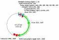
Rendering structure of FMDV IRES in an artificial transcript 181 nt long with energy of -20.90 kcal/mol as calculated by RNAeval using VARNA Java applet with some IRESite improvements (see VARNA modified by IRESite). Hold left mouse button to move structure parts, hold right mouse button to move whole structure, use mouse wheel to zoom. Right mouse-click opens a menu to export into JPG/SVG and many other options.
Remarks:
FMDV IRES domain3 with GUAA to GUAG mutation as shown in Fig. 8B
3.1.1. Enzymes used to characterize at least partially the 2D structure.
Enzyme or a combination of enzymes used in a single experiment with respective buffer:
ss_experiment_with_enzyme_id: 14
The temperature (in degrees of Celsia): 37
The enzymatic method used to determine the 2D structure: ribonuclease T1
Enzyme or a combination of enzymes used in a single experiment with respective buffer:
Version: 0
pH 7.50
Li+ [mM] 0
Na+ [mM] 50.00
K+ [mM] 300.00
Mg2+ [mM] 10.00
Ca2+ [mM] 0
Cl- [mM] 320.00
Tris [mM] 0
BSA [mM] 0
HEPES [mM] 0
EGTA [mM] 0
EDTA [mM] 0
cacodylate [mM] 50.00
Enzyme or a combination of enzymes used in a single experiment with respective buffer:
ss_experiment_with_enzyme_id: 16
The temperature (in degrees of Celsia): 20
The enzymatic method used to determine the 2D structure: ribonuclease T1
Enzyme or a combination of enzymes used in a single experiment with respective buffer:
Version: 0
pH 8.00
Li+ [mM] 0
Na+ [mM] 0
K+ [mM] 0
Mg2+ [mM] 0
Ca2+ [mM] 0
Cl- [mM] 0
Tris [mM] 10.00
BSA [mM] 0
HEPES [mM] 0
EGTA [mM] 0
EDTA [mM] 1.00
cacodylate [mM] 0
Enzyme or a combination of enzymes used in a single experiment with respective buffer:
ss_experiment_with_enzyme_id: 18
The temperature (in degrees of Celsia): 37
The enzymatic method used to determine the 2D structure: ribonuclease A
Enzyme or a combination of enzymes used in a single experiment with respective buffer:
Version: 0
pH 7.50
Li+ [mM] 0
Na+ [mM] 50.00
K+ [mM] 300.00
Mg2+ [mM] 10.00
Ca2+ [mM] 0
Cl- [mM] 320.00
Tris [mM] 0
BSA [mM] 0
HEPES [mM] 0
EGTA [mM] 0
EDTA [mM] 0
cacodylate [mM] 50.00
Enzyme or a combination of enzymes used in a single experiment with respective buffer:
ss_experiment_with_enzyme_id: 20
The temperature (in degrees of Celsia): 20
The enzymatic method used to determine the 2D structure: ribonuclease A
Enzyme or a combination of enzymes used in a single experiment with respective buffer:
Version: 0
pH 8.00
Li+ [mM] 0
Na+ [mM] 0
K+ [mM] 0
Mg2+ [mM] 0
Ca2+ [mM] 0
Cl- [mM] 0
Tris [mM] 10.00
BSA [mM] 0
HEPES [mM] 0
EGTA [mM] 0
EDTA [mM] 1.00
cacodylate [mM] 0
Enzyme or a combination of enzymes used in a single experiment with respective buffer:
ss_experiment_with_enzyme_id: 22
The temperature (in degrees of Celsia): 37
The enzymatic method used to determine the 2D structure: ribonuclease V1
Enzyme or a combination of enzymes used in a single experiment with respective buffer:
Version: 0
pH 7.50
Li+ [mM] 0
Na+ [mM] 50.00
K+ [mM] 300.00
Mg2+ [mM] 10.00
Ca2+ [mM] 0
Cl- [mM] 320.00
Tris [mM] 0
BSA [mM] 0
HEPES [mM] 0
EGTA [mM] 0
EDTA [mM] 0
cacodylate [mM] 50.00
3.1.2. Chemicals used to characterize at least partially the 2D structure.
Chemical reagent used with its respective buffer:
ss_experiment_with_chemical_id: 17
The temperature (in degrees of Celsia): 20
The chemical reagent used to determine the 2D structure: DMS
Chemical reagent used with its respective buffer:
Version: 0
pH 7.50
Li+ [mM] 0
Na+ [mM] 50.00
K+ [mM] 300.00
Mg2+ [mM] 10.00
Ca2+ [mM] 0
Cl- [mM] 320.00
Tris [mM] 0
BSA [mM] 0
HEPES [mM] 0
EGTA [mM] 0
EDTA [mM] 0
cacodylate [mM] 50.00
Chemical reagent used with its respective buffer:
ss_experiment_with_chemical_id: 18
The temperature (in degrees of Celsia): 20
The chemical reagent used to determine the 2D structure: DMS